Comparative Analysis of COVID - 19 Image Processing With X-Ray And CT Scan
1Amit Kumar Arora, 2Jyoti Parashar, 3Kamal Upreti, 4Balram Rao

Corresponding Author: Dr. Kamal Upreti
Email Id:kamalupreti1989@gmail.com
1Department of Computer Science and Engineering, IIT Madras, India
2,3Department of Computer Science & Engineering, ADGITM, Delhi, India
4Asia Pacific Institute of Management, New Delhi, India
Email Id:manshguide@gmail.com,
Email Id:jyoti.parashar123@gmail.com
Email Id:kamalupreti1989@gmail.com,
Email Id:balram.rao@asiapacific.edu
APFCB News Volume 2, Issue 1
General introduction
AI and its applications are among the most explored research regions. Over recent years, we have seen AI upsetting a wide range of clinical imaging, including chest XRay, ultrasound.
CT, fMRI, PET, as well as SPECT. Different AI-situated devices have to modernize clinical picture assessment and further create robotized picture interpretation. Modern medical imaging methods allow doctors to access data from a wide range of different sorts of research, including artificial intelligence. Deep learning, fuzzy sets, rough sets, doubtful analysis, multi-target development, swarm knowledge improvement, and machine learning are only few of the many areas of study that often make use of these elements. Patients are surveyed according to clinical groups using the delayed findings of these commonly used tests as a data point. By taking use of AI models' capacity to understand patterns and connections in clinical images, this new experiment in computer intelligence-driven configurations enhances tools for individualized infection evaluation. To do this, clinical images are used as the training data.
Prevalence of covid-19 disease
In the latter half of 2019, the SARS-COV2 (COVID-19) outbreak began in Wuhan City in China, and it eventually became a pandemic over the whole world. The pandemic caused by the COVID-19 virus has spread to more than 220 countries and territories throughout the world and has had an effect on every aspect of our day-to-day life. The quantities of contaminated cases passing despite everything increment significantly, which does not indicate a very well controlled circumstance; as of the 22nd of January in the year 2022, a total aggregate of 34,64,64,304 (55,85,224) contaminated (deceased) COVID-19 cases were accounted for all over the world [1]. The World Health Organization (WHO) has classified this illness as a global epidemic and has recommended a number of preventative actions in response to the growing severity of the pandemic. In the 21st century, Human COVIDS like as SARS-CoV and MERS-CoV have emerged out of the animal supplies-induced global pandemic, bringing with them a distressingly high mortality rate as well as morbidity. Infections caused by the COVID-19 virus seen in humans are classified under the Corona vitrine subgroup, which is part of the larger Corona viridae group. It was given the name COVID-19 owing to the presence of spikes in the pattern on the outside surface of the infection when seen under an electron microscope. Its RNA is a lone abandoned molecule with a diameter ranging from 80 to 120 nm, and the length of the nucleic material reaches varies between 26 and 32 Nucleotides [2]. In its most basic form, they may be broken down into four different genera known as, whereas -Cove and -Cove are mostly responsible for infecting vertebrates, -Cove and -Cove are primarily responsible for infecting avian species. HCoV-229E, in addition to HCoV-NL63 of -Coves, as well as HCoV-HKU1 and HCoV-OC43 of -Coves, demonstrates modest pathogenicity as well as mild respiratory side effects as a normal virus. This makes them one of the six defenseless human illnesses.
Figure 1 exhibits the total number of reported cases. Figure 2 represents the daily number of deaths reported up to 22nd January, 2022.
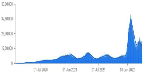
Figure 1- Number of active cases up to 22nd January, 2022 [1]
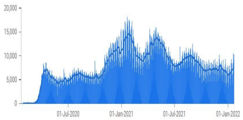
Figure 2- Number of deceased cases up to 22nd January, 2022 [1]
Characteristics of COVID-19
While the profoundly pathogenic infection predominantly influences individuals through respiratory droplets, disseminated via ecological contact [5, 6]. Unlike the various respiratory infections, other studies [7, 8] recommended that the SARS-CoV-2 communicated through the oral-fecal course. An ongoing investigation was done in ref. [9] on stool specimens of seventy-one individuals with COVID-19, 39 individuals +ve for fecal COVID-19 RNA, which supports the speculation that fecal-oral contamination could be an extra course for the extent of the infection. Generally, COVID- 19 manifestations are cough, fever, as well as fatigue [10–12].Various gastrointestinal indications showed in contaminated individuals like diarrhea, nausea, as well as deficiency of appetite [13, 14]. It is also important to note that the infection contamination could happen with no manifestation; asymptomatic people are a possible wellspring of infection transmission.
Consequently, stringent adherence to the climate, hand cleanliness, as well as contact segregation is needed to guarantee viral control. The ACE2 is the cell receptor for COVID-19 as well as various respiratory asperity condition viruses [15]. It acts a real job in the guideline of intestinal aggravation as well as the management of heart physiology. In any case, it has been accounted for that ACE2 is a cell-surface receptor for the infection, which works with the viral RNA section in the lung [16, 17].
Aspects of medical imaging modalities in the prognosis of COVID-19
The RT-PCR is usually employed being the determination of COVID-19. This biochemical response utilizes RNA rather than DNA as an initial step [8]. The reverse transcriptase chemical utilizes the RNA to generate a corresponding single-abandoned DNA in the reverse transcription measure. Then, at that point, the new integral single DNA is changed over into twofold abandoned DNA since it is utilized as a format for a PCR reaction [1, 2]. Even though numerous endeavors have been led to augment the number of PCR tests each day, this method experiences a few limits. The remarkable pace of false-negative recognized in individuals identifying COVID-19, the nonaccessibility of PCR reagent kits, and the generally lengthy timespan interaction of this technique [2]. Various more precise procedures or practices are expected to help determine COVID-19 infected individuals in such a setting. In this circumstance, clinical imaging methods assume a significant part in separating patients chosen from a frontline medical emergency and in the portrayal of pulmonary inclusion of COVID-19. The role of clinical imaging in battling the epidemic of COVID-19 and achieved outcomes of each imaging approach are explained.
COVID-19 exists in the form of various mutants, making epidemic circumstances around the globe. Thus, the clinical prognosis is not precise. However, different medical predictive techniques are in practice, viz. CXR, Computed Tomography, a furthermore non-radiation technique like ultrasound diagnostic method to augment the analytical techniques (for example, RT-PCR) to a definite restraint.
Chest X-Ray
Description: A typical X-ray utilizes an immovable tube that transmits X-rays in a single direction.
Working Principle: An electromagnetic wave of immense energy as well as shortened wavelength, which can send over the body.
Wavelength range: 10-9 to 10-12 meters and Energy range: 124 eV to 124 Kev.
Usage: To detect dislocations and fractures of bones as well as to detect cancers and Pneumonia.
Given the large number of medical infectious of COVID-19, the Chest X-ray method is possibly a convenient tool for identifying the infection because its vacancy correlated to CT tools. In a few nations like Spain, X-ray is the primary imaging procedure utilized for the prognosis of COVID-19 infected individuals [2].
The significant characteristics noticed in X-ray image individuals with COVID -19 were GGO as shown in figure 3, pulmonary nodules as well as interstitial variations [3, 4]. The Ground Glass Opacities were described as hazy regions by expanded lung thickness without obscuration of bronchial designs as well as vessels [18, 19].
Likely the rise of infected cases, the adoption of X-rays could perform a significant aspect. It may inhibit CT analysis, especially in the nations with restricted availability of the RT-PCR method. Nonetheless, in extremely medically infected, an RT- PCR test / Computed Tomographic films should be carried out [20, 21].
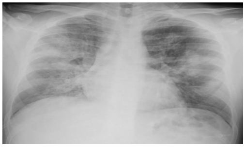
Figure 3- A sample of COVID-19 infected Chest X-ray image shows peripheral opacities in all pulmonary lobes.
Chest Computed Tomography
Description: Computed Tomographic films are associated with abundant X-ray measurements, collected from various angles to yield cross-sectional pictures of a particular region of a scanned body
Working Principle: A mechanized X-ray source that explodes narrow beams of X-rays as it pivots around the individual.
Usage:: Particularized images of abundant structures inside the body, containing the bones, internal organs, as well as blood vessels.
Under current medical involvement, Chest CT is contemplated as the best accurate tool for identifying COVID-19 disease [5]. Albeit various early prognosis tests of viral infectious like RT-PCR as well as biomarkers [6, 7]. The CT pictures give more signs of infection movement; furthermore, it is endorsed for asymptomatic individuals with - ve nucleic acid testing [8].
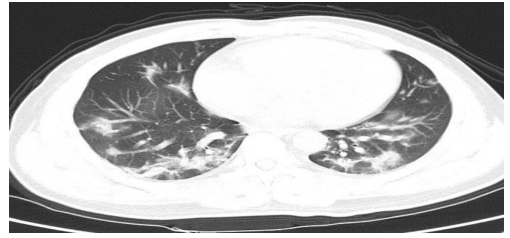
Indeed, affectability of chest Computed Tomography is better than RT-PCR in recognition of COVID-19 disease as exhibited in Fang et al. [9], as well as was illustrated in the analysis that enclosed 1014 individuals amidst +ve RT-PCR test that revealed an affectability of 97 percent of chest Computed Tomography in the identification of COVID-19 [3]. In general, CT identification is the crazy-paving pattern that resides in linear patterns overlapped on circumstances of Ground Glass Opacities [1, 2], as indicated as shown in figure 4. Various abnormal features were accomplished in CT pictures like pleural effusion, lung fibrosis, and airway transitions [3, 4].
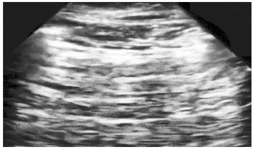
Figure 4- A sample of COVID-19 infected CT scan image shows the crazy-paving pattern
Chest Ultrasound
Description A piezoelectric crystal vibrates when an electric signal is enforced creating high-recurrence sound waves.
Working Principle Transducer transmits sound waves inside the body, gathers the reverberations that are reflected, and transfers them to PC, which makes the pictures.
Sound wave range 2 to 18 MHz
Usage To detect the organs damage, tissues, and other structures inside the body.
Figure 5- A sample of COVID-19 infected Ultrasound image
Researchers concentrated on X-ray and CT results for COVID-19 diagnosis to the greatest extent possible. Ultrasound images and their potential inputs were used to do some analysis that helped diagnose the illness. Ultrasound artefacts from the pleural surface and the chest wall are important to analyse for diagnosing lung infections. As shown in figure 5, the B lines artefacts, in their varying manifestations, are characteristic of COVID-19 in LUS images [5]. This is particularly true for the situation of intermediate asperity. The focus on this artefact as a specific lung infection lineage is growing. Broad bands of tissue in the lungs emerge at the pleural line and move forward in a coordinated manner through lung sliding, as described in [6,25].
Additional indicators of COVID-19 in LUS images include pleural line abnormalities. The average range separates the horizontal lines below the pleural lines in normal lungs. These pleural lines seem broken, uneven, and thickened to the person living with COVID-19 [22, 23].
The LUS method may be useful in determining the presence or absence of an active infection, and it certainly has the advantage of being less expensive [24]. Chest ultrasounds have limited use in medicine, being limited to detecting abnormalities in the lungs' periphery [7].
Medical Imaging Positioned on AI on the Prognosis of COVID-19
In the face of widespread worries over the COVID-19 pandemic, the majority of countries have adopted the policy of home isolation for infected persons who move through moderate or advanced phases of the disease. Nevertheless, using this strategy results in an extremely high mortality rate, which is proportional to the number of patients that are infected. The rapid worsening in patients' health brought on by COVID-19 at the emergency stage of the disease in the presence of very severe circumstances [8] may help to explain the rise in the mortality rate. In severe cases of respiratory failure, heart attack, or severe pneumonia, hospitalization is required as one of the next stages in the treatment process.
It is possible that clinical imaging technologies that include AI techniques will be vital in the future since they will provide patients with a quick and early diagnostic mechanism during times of severe illnesses. In addition, AI would be able to determine the major vulnerability instances that are associated with the quick progression of serious consequences for people who have COVID-19, which would make it possible to expand the treatment plan.
Conclusion
The findings that were presented and reported in the dissertation were the result analysis of radiological pictures to locate people who were infected with COVID-19. At the preliminary phase of the pandemic, simulation was carried out on a small number of the samples that were available in the repository. As time goes on, more photographs are added to the repositories as a result of the rising number of incidents that occur each day. An early detection of COVID-19 using Ultrasound pictures may
There by minimize the severity level and prevent the infection from spreading to other people. The approaches that were exhibited produced superior performance accuracy with big and balanced picture datasets in comparison to previous published studies. Neither overfitting nor under fitting will have any effect on the suggested models while they are being trained or tested. After receiving this first diagnosis, these processes could be of use to the doctors and radiologists in caring for COVID-19 infected patients.
[1] Google News: https://news.google.com/covid19/map?hl=en-IN&gl=IN&ceid= IN%3Aen. Accessed on 22nd January 2022.
[2] Zhu, N., Zhang, D., Wang, W., Li, X., Yang, B., Song, J., Zhao, X., Huang, B., Shi, W., Lu, R. and Niu, P., 2020, “A novel coronavirus from patients with pneumonia in China, 2019”, New England journal of medicine, 382, pp. 727-733.
[3] Yin, Y. and Wondering, R.G., 2018, “MERS, SARS and other coronaviruses as causes of pneumonia”, Respirology, 23(2), pp. 130-137
[4] 3D medical animation corona virus.jpg- Wikimedia Commons. Wikimedia Commonsn.d.https://commons.wikimedia.org/wiki/File:3D_medical_animation corona_viru.jpg. Accessed on 8th August, 2021.
[5] Liu, J., Liao, X., Qian, S., Yuan, J., Wang, F., Liu, Y., Wang, Z., Wang, F.S., Liu, L. and Zhang, Z., 2020, “Community transmission of severe acute respiratory syndrome coronavirus 2, Shenzhen, China, 2020”, Emerging infectious diseases, 26(6), pp. 1320.
[6] Jiang F, Deng L, Zhang L, Cai Y, Cheung CW, and Xia Z, 2020, “Review of the clinical characteristics of coronavirus disease 2019 (COVID-19)”, Journal of General Internal Medicine, 35, pp. 1545-1549
[7] Yeo, C., Kaushal, S. and Yeo, D., 2020, “Enteric involvement of coronaviruses: is faucal–oral transmission of SARS-CoV-2 possible?” The lancet Gastroenterology & hepatology, 5(4), pp. 335-337.
[8] GU, J., Han, B. and Wang, J., 2020, “COVID-19: gastrointestinal manifestations and potential fecal–oral transmission”, Gastroenterology, 158(6), pp. 1518-1519.
[9] Xu, Y., Li, X., Zhu, B., Liang, H., Fang, C., Gong, Y., Guo, Q., Sun, X., Zhao, D., Shen, J. and Zhang, H., 2020, “Characteristics of pediatric SARS-CoV-2 infection and potential evidence for persistent fecal viral shedding”, Nature medicine, 26(4), pp. 502-505
[10] Fu, L., Wang, B., Yuan, T., Chen, X., Ao, Y., Fitzpatrick, T., Li, P., Zhou, Y., Lin, Y.F., Duan, Q. and Luo, G., 2020, “Clinical characteristics of coronavirus disease 2019 (COVID-19) in China: a systematic review and meta-analysis”, Journal of Infection, 80 (6), pp. 656-665.
[11] Pernazza, A., Mancini, M., Rullo, E., Bassi, M., De Giacomo, T., Della Rocca, C. and d’Amati, G., 2020, “Early histologic findings of pulmonary SARS-CoV-2 infection detected in a surgical specimen”, Virchows Archive, 477(5), pp. 743-748
[12] Zhang, R., Ouyang, H., Fu, L., Wang, S., Han, J., Huang, K., Jia, M., Song, Q. and Fu, Z., 2020, “CT features of SARS-CoV-2 pneumonia according to clinical presentation: a retrospective analysis of 120 consecutive patients from Wuhan city”, European radiology, 30(8), pp. 4417-4426
[13] Cholankeril, G., Podboy, A., Aivaliotis, V.I., Tarlow, B., Pham, E.A., Spencer, S., Kim, D., Hsing, A. and Ahmed, A., 2020, “High Prevalence of Concurrent Gastrointestinal Manifestations in Patients with SARS-CoV-2: Early Experience from California”, Gastroenterology, 159(2), pp. 775-777
[14] Xiao, F., Tang, M., Zheng, X., Liu, Y., Li, X. and Shan, H., 2020, “Evidence for gastrointestinal infection of SARS-COV-2” Gastroenterology, 158(6), pp. 1831-1833.
[15] Zhang, H., Penninger, J.M., Li, Y., Zhong, N. and Slutsky, A.S., 2020, “Angiotensinconverting enzyme 2 (ACE2) as a SARS-CoV-2 receptor: molecular mechanisms and potential therapeutic target”, Intensive care medicine, 46(4), pp. 586-590.
[16] Jia, H.P., Look, D.C., Shi, L., Hickey, M., Pewe, L., Netland, J., Farzan, M., WohlfordLenane, C., Perlman, S. and McCray Jr, P.B., 2005, “ACE2 receptor expression and severe acute respiratory syndrome coronavirus infection depend on differentiation of human airway epithelia”,Journal of virology, 79(23), pp. 14614-14621.
[17] Misra, D.P., Agarwal, V., Gasparyan, A.Y. and Zimba, O., 2020, “Rheumatologists’ perspective on coronavirus disease 19 (COVID-19) and potential therapeutic targets”, Clinical rheumatology, 39(7), pp. 2055-2062.
[18] Shreshth Tuli, Shikhar Tuli, Gurleen Wander, Praneet Wander, SukhpalSingh Gill, Schahram Dustdar, Rizos Sakellariou, Omer Rana, Nextgeneration technologies for smart healthcare: challenges, vision, model,trends and future directions, Internet Technol. Lett. 3 (2) (2020) e145.
[19] Maxwell W. Libbrecht, William Stafford Noble, Machine learning appli-cations in genetics and genomics, Nat. Rev. Genet. 16 (6) (2015)321–332.
[20] Mohammad Jamshidi, Lalbakhsh Ali, Jakub Talla, Zden?ek Peroutka,Farimah Hadjilooei, Pedram Lalbakhsh, Morteza Jamshidi, Luigi LaSpada, Mirhamed Mirmozafari, Mojgan Dehghani, et al., Artificialintelligence and covid- 19: deep learning approaches for diagnosis andtreatment, IEEE Access 8 (2020) 109581– 109595.
[21] A. Singh, D. Singh, K. Upreti, V. Sharma, B. Singh Rathore, and J. Raikwal, “Investigating New Patterns in Symptoms of COVID-19 Patients by Association Rule Mining (ARM),” Aug. 25, 2022. https://doi.org/10.13052/jmm1550-4646.1911 (accessed Mar. 05, 2023).
[22] Pradeep Kumar Dabla, Kamal Upreti, Divakar Singh, Anju Singh, Jitender Sharma, Aashima Dabas, Damien Gruson, Bernard Gouget, Sergio Bernardini, Evgenija Homsak & Sanja Stankovic (2022) Target association rule mining to explore novel paediatric illness patterns in emergency settings, Scandinavian Journal of Clinical and Laboratory Investigation, 82:7-8, 595-600, DOI: 10.1080/00365513.2022.2148121.
[23] Upreti, K. et al. (2023). IoT-Based Control System to Measure, Analyze, and Track Basic Vital Indicators in Patient Healthcare Monitoring System. In: Shaw, R.N., Paprzycki, M., Ghosh, A. (eds) Advanced Communication and Intelligent Systems. ICACIS 2022. Communications in Computer and Information Science, vol 1749. Springer, Cham. https://doi.org/10.1007/978-3-031-25088-0_63.
[24] Upreti, K. et al. (2023). A Deep Convolution Network-Based Pneumonia Identification from Thoracic X-Ray Imagery Scans. In: Nagar, A.K., Singh Jat, D., Mishra, D.K., Joshi, A. (eds) Intelligent Sustainable Systems. Lecture Notes in Networks and Systems, vol 578. Springer, Singapore. https:/ /doi.org/10.1007/978-981-19-7660- 5_64
[25] Syed MH, Upreti K, Nasir MS, Alam MS, Kumar Sharma A. Addressing image and Poisson noise deconvolution problem using deep learning approaches. Computational Intelligence. 2022;1-15. DOI: 10.1111/coin.12510